FINITE ELEMENT MESHING
OF HUMAN BONES FROM MRI RAW DATA
Finite element modeling of human bones is quite useful in
biomechanical simulations. In the present project work a new
approach is developed to make FE model of bones from MRI
scan data. Developed technique is modification over
conventionally used technique. In conventional technique
solid modeling process is essential before getting finite
element model from the MRI scan data. In present project
work this necessity is eliminated and process time and steps
are shortened.
Conventional process for finite element meshing from MRI
scan data requires two intermediate steps first interior and
exterior contour point extraction of bones and second solid
modeling from contour data extracted. In the methodology
developed and implemented in present work solid generation
process is completely eliminated and finite element meshes
can be developed directly from the contours extracted. In
the first part of project work a user interface is developed
to extract contour point data from raw MRI scan data.
Process of getting contour data involves getting scan images
from raw data, getting bone region from scan images and
finally getting contour data extraction. Contour data are
obtained using some image processing techniques.
In second part of project work another user interface in the
same language is developed to make FE model from contour
data extracted. This process involved mapping of contours on
unit rectangle to get quadrilateral mesh within each contour
and fitting solid element in between two consecutive
contours.
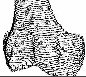
Lofted contour points of the femur
(Chawla A. et al., 2006)
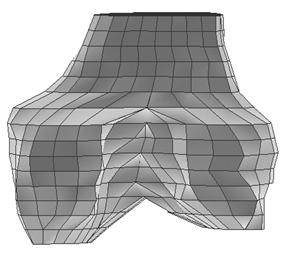
Meshed model of femur end from manual
meshing (Chawla A. et
al., 2006)